
Summary
Da Vinci robot was used for cardiothoracic surgery at PA Hospital for the first time in July 2026.
The adopted surgical technique using ICG dipped coil and infrared light is paving the way for new applications in chest and lung surgery.
PA Hospital’s first patient to receive thoracic surgery with the assistance of the robot has paved the way for new applications within the cardiothoracic surgery team.
Thanks to the agility of the robot, previously difficult-to-reach lesions in the lung or the chest cavity are now treatable with easier access and reduced recovery time through this less invasive surgical option.
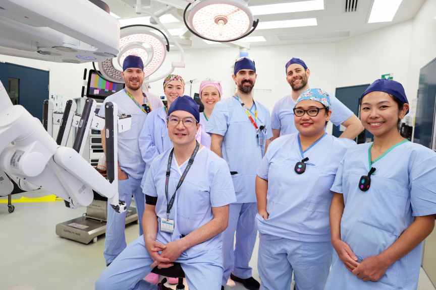
While the Da Vinci robot has been in use for multiple specialties at PAH for over a decade, cardiothoracic surgeon, Dr Chris Cole said the July 2026 surgery was the first time thoracic surgery has been performed using the robot at PAH, and only the second time in a Queensland public hospital after the Gold Coast University Hospital.
"The advantage of the robot for chest surgery is better visualisation and access for hard to reach corners of the chest, without making a big cut between the ribs, or using limited rigid instruments of previous keyhole techniques."
“Those techniques offered limited angles and restricted effective access to multiple locations in the lungs,” Dr Cole said.
In addition to the better instruments of the robot, the robot allows for "seeing" with near infrared light.
“The problem with small lung lesions is that it is difficult to feel the lumps in order to find and remove them.” Dr Cole said. "So for this patient, our radiology team marked the lesions in the lungs with ICG, which glows green under near infrared light."
To find small lesions in the lungs, surgeons previously made an incision between the ribs, and spread them apart so that hands could access the chest to feel around for the lumps.
Introducing the robot to thoracic surgery involved training the broader surgical team and introducing a new Indocyanine green (ICG) dipped coil technique through Radiology.
Radiologist Dr David Chang said the new technique involved the placement of special coils coated with ICG which highlights the nodule and coil when the robot uses a special infrared light.
“Under CT guidance, we can insert a needle within 4-5mm of the lesion which allows us to put a coil next to the lesion and run a lead like a fishing line to the surface of the lung.” Dr Chang said.
“I leave a small ball on the surface of the lung like a fishing bobber so that the surgeon can see where the nodule is with the robot.
“The coil mixed with the green allows the surgeon using the robot to see past the surface of the lung using the special infrared light to locate the nodule precisely.”
While this was not Dr Cole’s first use of the robot for thoracic surgery, it was a first for the PAH surgical team alongside the introduction of the ICG dipped coil which will revolutionise early intervention for future thoracic surgeries.
“This was a momentous first step for the team heading into the new frontier of robotic thoracic surgery,” Dr Cole said.
“The less trauma the patient experiences during the course of that treatment, the better their recovery each time. That’s the goal.”
The expanded lung cancer screening program is coming soon for earlier detection of lung nodules. This will enable earlier intervention and greater preservation of normal lung with this technique; providing a minimally invasive option for nodule removal when it is too small to biopsy.